Teilnahmebedingungen für das Gewinnspiel “Brain Boost Event 2025 – Gewinne 2x Teilnahmeplätze für dich und deinen Lieblingskollegen in Seefeld”
Teilnahmebedingungen:


How often do you encounter Molar Incisor Hypomineralization (MIH) in your practice? While often overlooked, MIH affects 13-14 % of children worldwide and can have a major impact on quality of life. Learn more about this common, serious condition and what you can do to help.
Molar Incisor Hypomineralization (MIH) is a very common developmental dental condition – so common, in fact, that it’s estimated to affect 13-14% of children worldwide. Despite this, many clinicians have trouble diagnosing and managing the problem. With this in mind, it’s critical to develop a deeper understanding of the condition, was well as review prevention and treatment strategies, in order to address the problem as early as possible.
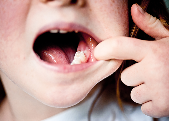
Known by many names including “chalky teeth” MIH is defined as a qualitative developmental defect of enamel, affecting at least one first permanent molar (with or without involvement of the incisors). Depending on the severity of the condition, hypomineralized enamel can be soft, porous, or even resemble cheese. It can vary both in size and in color from white to yellow or brown. However, there is always a sharp demarcation between affected and sound enamel. MIH can also progress to and present as posteruptive enamel breakdown: the enamel has grown so brittle that, under chewing forces or during eruption through gingiva, the tooth has started to break. This can lead to exposed dentin, leaving the tooth vulnerable to caries. Affected molars present along a spectrum of severity and defect extension, from barely noticeable
opacities to severe destruction of enamel and extreme hypersensitivity, leading to discomfort and functional issues.
With respect to hypersensitivity, children often report that intake of hot and cold or sweet food, toothbrushing, and even air flow cause pain. Hypersensitivity already occurs during eruption of the molar and therefore needs an early treatment.
MIH is a disease that occurs worldwide. However, the prevalence of MIH varies greatly in different parts of the world (2.8 – 40.2%) due to a lack of standardized tools for diagnosis and recording.1 In any case, it’s estimated that MIH affects one in eight children worldwide, so it’s very likely you’ll see the condition in your practice.
While MIH was first reported in the 1970s, its true etiology is still unclear. Several research groups are discussing a multifactorial cause as most likely.2 It seems to be certain that tooth development is disturbed in the first few years of life caused by antibiotics, infection, disease, or problems during pregnancy.3
If left untreated, MIH can have a major impact on the child’s quality of life and overall health. Due to associated sensitivity and risk of caries, children can experience discomfort and pain, and difficulty eating, drinking, and speaking. This, along with the esthetic impact, can have long-term social and psychological effects. Because knowledge of MIH is still developing and it can’t be identified until the tooth erupts, it’s easy to overlook or misdiagnose the situation as carious. However, the earlier the condition is caught, the earlier prevention and treatment can begin.
In an effort to simplify assessment and treatment selection, the European Academy of Pediatric Dentistry (EAPD) has developed a grading system for MIH-affected teeth.4 Using this system, affected teeth can be assessed on a scale of mild to severe. Mild cases feature teeth with limited opacities without any loss of tooth structure, occasional hypersensitivity to external stimuli (except tooth brushing), and only mild esthetic concerns due to incisor discoloration. On the other end of the spectrum, severe cases are defined by demarcated opacities with enamel erosion, caries, extreme hypersensitivity, and severe esthetic concerns on the incisors that could have an impact on the child’s self-esteem.
Before this system, previous classification systems did not include any therapeutic guidance. The new system addresses this issue with a classification index, “MIH Treatment Need Index (MIH-TNI),” and associated therapy concept that gives clear guidance on how to treat MIH-affected teeth in different stages of severity.5,6 The system classifies four severity codes:
• MIH-TNI 1: MIH without hypersensitivity or breakdown
• MIH-TNI 2: MIH without hypersensitivity, but with breakdown
o 2a: <1/3 defect extension
o 2b: 1/3 – 2/3 defect extension
o 2c: >2/3 defect extension or/and defect close to the pulp or extraction or atypical restoration
• MIH-TNI 3: MIH with hypersensitivity, but without defect
• MIH-TNI 4: MIH with hypersensitivity and defect
o 4a: <1/3 defect extension
o 4b: 1/3 – 2/3 defect extension
o 4c: >2/3 defect extension or/and defect close to the pulp or extraction or atypical restoration
Using this system, clinicians can identify all problems and begin planning treatment with the appropriate therapy options.

Treating MIH can be challenging for a number of reasons, including the child’s age, compliance, post-eruptive breakdown, and hypersensitivity but as mentioned above, early diagnosis is key to effective and conservative management.
Due to the soft, porous enamel, post-eruptive enamel breakdown is likely. The breakdown exposes sensitive dentin and makes the tooth more susceptible to caries, which can lead to crown destruction, atypical restorations or even tooth loss. Exposed dentin can also lead to hypersensitivity and pain which can limit treatment options, as achieving adequate pain control can be challenging. Plus, bonding to this porous enamel can be difficult, frequently resulting in loss of fillings or repeated treatment. Together, all these factors can reduce kids’ compliance due to anxiety about treatment. And to top it off, parents must understand the gravity of the situation, cooperate with treatment suggestions,
and help uphold hygiene recommendations for treatment to be effective in the long run. With all of this in mind, how do you go about treatment?

Depending on the severity of the MIH, different therapeutic approaches should be considered: from intensive prophylaxis to restorative measures to extraction.7 Regardless of the severity of the MIH, all affected children should be closely cared for in an intensive prophylaxis program (see graph above).
Prevention and management should start as soon as MIH teeth erupt, as they are prone to post-eruptive breakdown and caries. This effort begins with educating parents on proper dietary and hygiene habits, the importance of in-office fluoridated treatment with a calcium source, as well as encouraging the use of fluoridated toothpaste to reduce the risk of caries – but doesn’t stop there.
Pit and fissure sealants serve a critical role in prevention, as they help reduce sensitivity and protect MIH-affected teeth from caries and further breakdown until the tooth has fully erupted. Resin-based sealants are very popular; however, they can only be placed on fully erupted molars where the enamel surface is intact and moisture control is adequate. An etching step or adhesive is needed to improve bonding on the porous, less mineralized enamel.8 In cases where the tooth is not fully erupted and moisture control is difficult, using a moisture tolerant, low-viscosity glass ionomer cement can be helpful.
As mentioned earlier, compared to filling therapy on healthy teeth, the treatment of MIH molars presents special challenges for the practitioner. These include the age of the patient, the severity and extent of the loss of enamel as well as the extent of the hypersensitivity of the affected teeth.9
Glass ionomer restorative materials (GIs) present an incredible opportunity to protect MIH-affected teeth while increasing patient compliance and comfort. GIs are already most dental professionals first choice for an interim restorative material. They’re ideal as an initial and temporary covering of erupting teeth to avoid further loss of enamel. GIs don’t require an etching step or an adhesive system, simplifying handling, facilitating application, and reducing time in the chair, which is particularly beneficial when patient cooperation is limited. And they don’t need additional conditioning steps as they chemically bond to natural dentin. Beyond simplifying the procedure, GIs actively protect teeth. Not only do they contain ions that support natural tooth structure, but they also release and absorb fluoride – providing a boost of protection for high-risk patients, such as those with MIH.
Prefabricated crowns
Prefabricated stainless-steel crowns work well as a long-term temporary solution. In cases of massive loss of enamel or hypersensitivity in young children, they can be used shortly after tooth eruption to preserve the tooth until a definitive indirect restoration is possible, or until the optimal time of extraction. The procedure is not very technique sensitive, and the stainless-steel crown can prevent further loss of enamel and protect remaining tooth structure.10
In certain cases, temporary treatment may not be an option, indicating more permanent solutions:
Direct restorations: Composites Due to their properties, composites are an ideal material to restore permanently MIH-affected teeth. These fillings can be placed in a minimally invasive manner, helping preserve healthy enamel. And when selecting an adhesive, studies have shown that self-etch adhesives produce significantly better adhesion values on MIH teeth than total-etch systems and are, therefore an at least equivalent, if not superior, option.11,12
Indirect restorations:
A composite-reinforced ceramic is a suitable material for onlays, overlays or (partial) crowns – and a long-lasting treatment option with a good long-term prognosis.13
Extraction
If the long-term prognosis of the MIH tooth is uncertain even after considering all available treatment options, extraction may be the best option.8 This last option can be considered if the tooth is affected by severe posteruptive enamel breakdown, has been severely damaged by caries, or pathological periapical processes are in progress.
MIH is a common, serious condition that affects the quality of life of many children worldwide. However, a combination of early diagnosis and early intervention with the right materials, like GIs, can help you set your patients up for strong smiles – throughout childhood and into the future.
References
1. Schwendicke F, Elhennawy K, Reda S, Bekes K, Manton DJ, Krois J: Global burden of molar incisor hypomineralization. Journal of dentistry 2018, 68:10-18.
2. Silva MJ, Scurrah KJ, Craig JM, Manton DJ, Kilpatrick N: Etiology of molar incisor hypomineralization–a systematic review. Community dentistry and oral epidemiology 2016, 44(4):342-353.
3. Laisi S, Ess A, Sahlberg C, Arvio P, Lukinmaa P-L, Alaluusua S: Amoxicillin may cause molar incisor hypomineralization. Journal of dental research 2009, 88(2):132-136.
4. Lygidakis N, Wong F, Jälevik B, Vierrou A, Alaluusua S, Espelid I: Best Clinical Practice Guidance for clinicians dealing with children presenting with Molar-Incisor-Hypomineralisation (MIH). European Archives of Paediatric Dentistry 2010, 11(2):75-81.
5. Bekes K, Krämer N, Van Waes H, Steffen R (2016) The Wuerzburg MIH concept: Part 2. The therapy plan. Oralprophylaxe & Kinderzahnheilkunde 38:165-171-175
6. Steffen R, Krämer N, Bekes K (2017) The Wuerzburg, MIH concept: The MIH treatment need index (MIH TNI): A new index to assess and plan the treatment in patients with molar incisor hypomineralization (MIH). Eur Arch Paediatr Dent 18:355-361
7. Bekes K: Molar incisor hypomineralization: Springer; 2020
8. Lygidakis N, Dimou G, Stamataki E: Retention of fissure sealants using two different methods of application in teeth with hypomineralised molars (MIH): a 4 year clinical study. European Archives of Paediatric Dentistry 2009, 10(4):223-226.
9. Elhennawy K, Schwendicke F: Managing molar-incisor hypomineralization: a systematic review. Journal of dentistry 2016, 55:16-24.
10. Bekes K: Neue Erkenntnisse bei der Therapie der Molaren-Inzisiven-Hypomineralisation (MIH) (New finding in the therapy of molar incisor hypomineralization (MIH) Plaque N Care, online, 202
11. Krämer N, Khac N-HNB, Lücker S, Stachniss V, Frankenberger R: Bonding strategies for MIH-affected enamel and dentin. Dental Materials 2018, 34(2):331-340.
12. 16. Lagarde M, Vennat E, Attal Jp, Dursun E: Strategies to optimize bonding of adhesive materials to molar‐incisor hypomineralization‐affected enamel: A systematic review. International journal of paediatric dentistry 2020, 30(4):405-420.
13. Davidovich E, Dagon S, Tamari I, Etinger M, Mijiritsky E: An innovative treatment approach using digital workflow and CAD-CAM part 2: The restoration of molar incisor hypomineralization in children. International journal of environmental research and public health 2020, 17(5):1499.

How do you motivate your patients? Discover how caries risk assessments and motivational interviewing tactics can help you connect with…

Caries is a complicated multifactorial disease. In this two-part series, explore how caries risk assessments can help improve evaluation and…